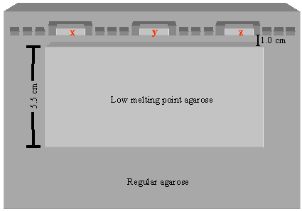
(a) Using a scalpel, join adjacent comb-made wells to form three slot-wells. Place a thin layer of LMP agarose in the bottom of each slot- well and allow the agarose to solidify. Insert blocks x, y, and z into the slot-wells. Place the PFGE lambda ladder into wells flanking the slot-wells as shown. Be sure to push x, y, and z to the leading edge of their respective slot-wells. Seal all wells with LMP agarose. Using a ruler as a guide, use a scalpel to cut out a large block of agarose from the center of the gel (see diagram). Fill the resulting space with 1% LMP agarose, and allow the LMP agarose to solidify. Run the gel using the following parameters: run time = 18 hr, initial switch time = 3 sec, final switch time = 5 sec, included angle = 120°, volts/cm = 6.0, buffer temperature = 12°C.
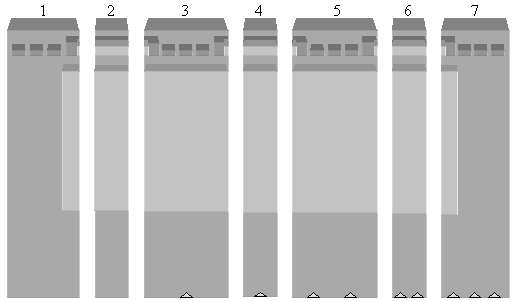
(b) Cutting the gel. After electrophoresis, divide the gel into seven pieces as shown above. The pieces are referred to as 1-7 from left to right. Cut notches into the bottom of pieces 3-7 as shown. Notches facilitate rapid reconstruction of the gel. Stain the odd-numbered pieces with ethidium bromide. Do not stain the even-numbered pieces as these contain genomic DNA which may be used in subsequent ligation reactions.
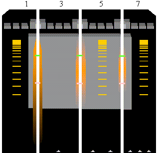
(c) Align the stained (odd-numbered) gel pieces and examine on a UV light box as shown. Using a scalpel, make incisions to mark the 125 kb (pink lines) and 350 kb (green lines) borders.
(d) Reconstruct the gel by placing the unstained even-numbered pieces between the stained odd-numbered pieces. Use the notches at the bottom of pieces 3-7 to facilitate proper assembly of the gel. Extend the incisions at 125 kb and 350 kb on each odd-numbered gel piece into adjacent even-numbered gel pieces (green and pink lines).
Note: The center gel piece should never be exposed to UV light from the light box as this can break the DNA!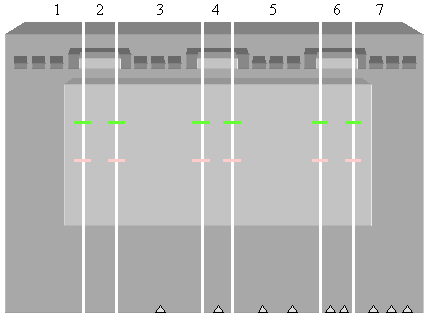
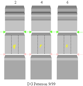
(e) Using a scalpel or razor blade, connect the appropriate incisions on each even-numbered gel piece as shown. The resulting center blocks containing DNA between 125 and 350 kb are referred to as q, r, and s, respectively. Subsequently, cut each of the three center blocks longitudinally to produce three blocks of roughly equal size (dotted lines). Place the three q blocks in one centrifuge tube, the three r blocks in a second tube, and the three s blocks in a third tube. The DNA in these blocks may be suitable for ligation.